|
I. Interface
Between a Ferromagnet and an Antiferromagnet – Exchange Bias System
We are using polarized neutron reflectivity and resonant magnetic x-ray
scattering to study the details of the depth dependence of the magnetization
and the lateral domain structure in the vicinity of the interfaces in compound
magnetic structures. The experiments are carried out at the Los Alamos
Laboratory Manuel Lujan Neutron scattering facility, the Spallation Neutron
Source at Oak Ridge National Laboratory, the Advanced Light Source at Lawrence
Berkeley National Laboratory, and the National Synchrotron Light Source at
Brookhaven National Laboraotry.
The systems being studied include ferromagnetic films deposited on
antiferromagnets (so-called "exchange-bias" systems), multilayers
exhibiting the giant magnetoresistance (GMR) effect or spin-valve systems,
ferromagnet/semiconductor interfaces (so-called "spin injection
systems"), magnetic nanodot arrays deposited on solid substrates, and hole
arrays in magnetic films.
Issues we are interested in include the correlation between interfacial
roughness and magnetic domain structure and coercive fields; the origin and
location of the so-called "uncompensated spins" in an antiferromagnet
in contact with a ferromagnet and their domain structure and its relation to
the domain structure in the ferromagnet as a function of applied magnetic
field; the magnetic domain structure in films with periodic microscopic arrays
of holes; the so-called vortex structure in magnetic nanodots; how the spin
structures of nanoparticles differ from those of the corresponding bulk
structures; the dynamics of how superparamagnetic moments fluctuate in magnetic
nanopartices as studied by coherent magnetic x-ray scattering; the depth
profile of spins injected into a semiconductor from a ferromagnet, etc.
We have developed a theoretical formalism for efficiently
calculating resonant magnetic scattering from magnetic interfaces with
roughness and domain structures [see the link ], which we use in
analyzing our magnetic x-ray scattering data.
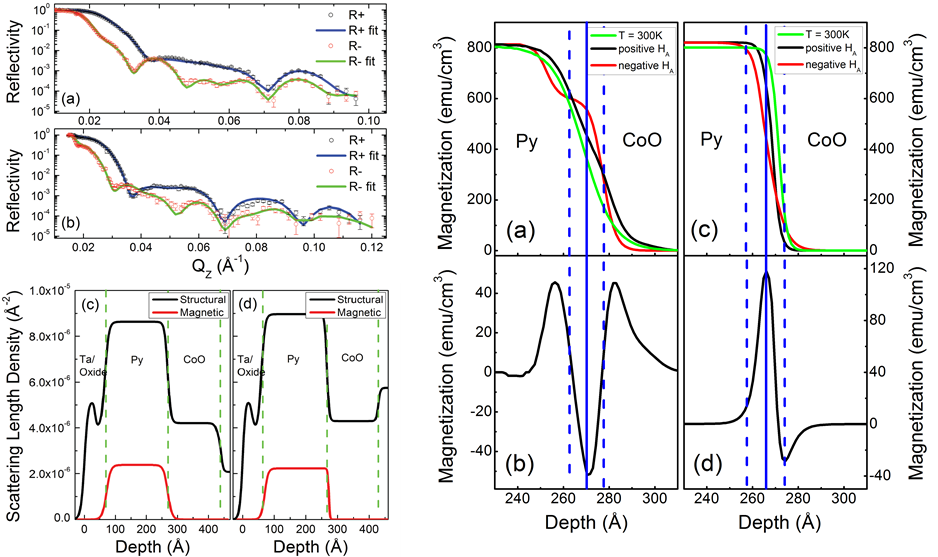
Figure.
(Left) Polarized neutron reflectivity of polycrystalline and (111) – epitaxial film
of permalloy/CoO exchange bias bilayer above Neel temperature. (Right) Magnetic
density profile at the interface and the pinned moments extracted from neutron
reflectivity at biased state.
II. Domain
Walls Fluctuation in Antiferromagnetic Dysprosium
We
use resonant magnetic X-ray scattering with coherent X-rays to observe magnetic
“speckles” from spin systems. Rare element dysprosium has a hexagonal close
packed structure and has a spiral antiferromagnetic structure between its Curie
temperature (~85K) and Neel temperature (~180K). With resonant soft X-ray, we
can reach the M-edge of dysprosium and obtain pure magnetic speckles at the
(0,0,Qm) diffraction peak. We used X-ray photon correlation spectroscopy (XPCS)
to study the dynamics of antiferromagnetic domain walls. We show that the
domains
of
a spiral antiferromagnet enter a jammed state at the onset of long-range order.
The slow thermal fluctuations of the domain walls exhibit a compressed
exponential relaxation with an exponent of 1.5 found in a wide variety of
solid-like jammed systems and can be qualitatively explained in terms of stress
release in a stressed network. As the temperature decreases, the energy barrier
for uctuations becomes large enough to arrest further domain wall uctuations
due to increase in exchange energy, and the domains freeze into a spatial configuration
within 10 K of the Neel temperature. The relaxation times follow the
Vogel-Fulcher law as observed in polymers, glasses and colloids thereby
indicating that the dynamics of domain walls in an ordered antiferromagnet exhibit
some of the universal features associated with glassy behavior.
The
experiment was carried out at beamline 12.0.2.2 at Advanced Light Source,
Lawrence Berkeley National Laboratory.
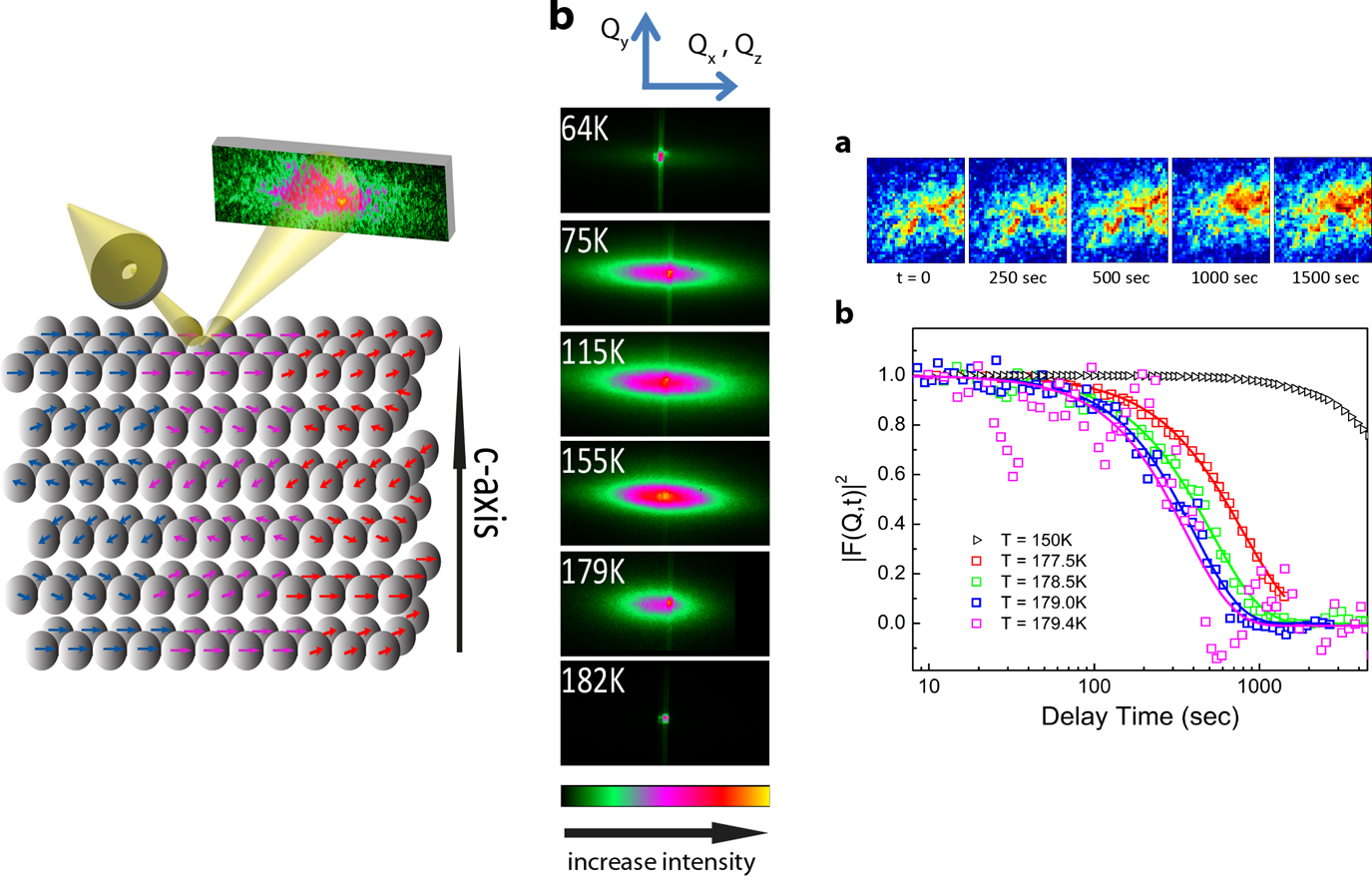
Figure.
(Left) Schematic diagram of spiral structure along the c-axis in dysprosium. Domains
are formed due to different charalities or phase slips. (Middle) Evolution of
magnetic diffraction peak as a function of temperature. Below Curie temperature
and above Neel temperature, only charge reflection peaks are seen. (Right)
Evolution of speckle pattern with time. The autocorrelation function shows a
strong dependence as a function of temperature.
|